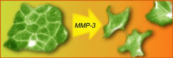
 Mutations in the genomes of the tumor cell long thought to cause cancer is negated by QED induced ionizing radiation produced as the MMP-3 enzyme disorganizes the basement membrane of epithelial tissues
Background Epithelial tissue forming the outer layers of the skin protect exterior surface of the body, but also provide protection for hollow organs and glands including the breast, prostate, colon, and lung from body fluids. Epithelial tissue is organized by a submicron thick < 100 nm basement membrane (BM) that provides the structural scaffold template for the extracellular matrix (ECM). Breakdown of the BM is associated with the spread of tumors, e.g., loss of integrity of the BM in mice is known to cause tumors. See http://www.lbl.gov/Science-Articles/Archive/LSD-cancer-f …
Loss of integrity in the ECM is triggered by enzymes called matrix metalloproteinases (MMPs). Breast tumors in particular are known to have an increased amount of MMPs. Indeed, MMPs induce the epithelial-mesenchymal transition (EMT) that disorganize the BM and allows the dissociated epithelial tissue to move through the body. In breast cancer, EMT allows tumor cells more mobility to penetrate barriers like the walls of lymph and blood vessels, facilitating metastasis, e.g., the MMP-3 enzyme causes normal cells to produce a protein called Rac1b that is found only in cancers. Currently, Rac1b is thought to stimulate the production of highly reactive oxygen species (ROS) molecules leading to cancer by damaging the DNA. Ibid
Problem and Hypothesis
The problem with epithelial tissue as the source of DNA damage is that the protein Rac1b lacks a mechanism to produce energy of at least 5 eV from which the ROS of peroxide and hydroxyl radicals form to damage the DNA. The ROS can only be produced by ionizing radiation > 5 eV at ultraviolet (UV) levels or beyond.
But what is the mechanism of ionizing radiation?
Certainly, there are no UV lasers in body fluids. The hypothesis may therefore be made that epithelial tissue during disorganization somehow emits low level ionizing radiation.
Emission of Ionizing Radiation by Submicron Entities
Cancer research is only beginning to recognize the remarkable fact that submicron entities present in body fluids emit low-level ionizing radiation. Over the past few decades, this fact has been supported by experimental evidence that shows submicron entities comprising nanoparticles (NPs) of natural and man-made materials cause DNA damage. Remarkably, the NPs alone – without lasers – somehow induce DNA damage in body fluids. See http://www.nanoqed.org/ at “DNA damage by NPs”, 2009 and “DNA damage by signaling,” 2010.
QED induced EM radiation
Recently, the theory of QED induced EM radiation was proposed to explain the remarkable fact that NPs alone cause DNA damage. QED stands for quantum electrodynamics and EM for electromagnetic. Ibid. By this theory, quantum mechanics forbids atoms in submicron NPs to have specific heat. This may be understood from the fact the thermal energy of the atom given by the Einstein-Hopf relation depends on wavelength under the constraint that only wavelengths < 1 micron are allowed to “fit inside” submicron entities. But quantum mechanics only allows submicron wavelengths to be populated at temperatures greater than about 6000 K. At ambient temperature, therefore, the heat capacity of submicron entities is “frozen out”, and so NPs cannot conserve absorbed EM energy by an increase in temperature. Absent UV lasers, NPs in body fluids absorb EM energy from colliding water molecules. Conservation then proceeds by the QED induced frequency up-conversion of absorbed EM collision energy to the fundamental resonance of the NP. Typically, ionizing QED radiation is emitted at UV or higher levels thereby explaining how NPs alone produce the ROS that damages DNA. Specifically, ionizing radiation > 5 eV necessary for forming ROS is produced for NPs < 100 nm. Ibid
Epithelial cell induced DNA damage
Epithelial tissue like NPs induce DNA damage provided submicron entities are produced upon disorganization by MMPs. QED induced radiation is emitted if the size of the biological entity is < 100 nm and has a refractive index greater than the surroundings. For biological materials, the index is about 1.5 > 1.33 for water. But epithelial cells themselves are not submicron and at 10-100 microns in the disorganized state do not emit ionizing radiation. However, ionizing radiation is emitted from the < 100 nm BM upon disorganization of epithelial tissue by MMPs.
By the theory of QED induced radiations, the disorganization of the BM by MMP-3 produces the ionizing radiation that forms the ROS that in turn damage the DNA. Current thought that the Rac1b protein itself damages the DNA is therefore placed in question. Indeed, the Rac1b protein is a cancer marker only because it forms in the process of ionizing radiation be produced by the disorganized BM. Nevertheless, Rac1b as a small protein is a submicron entity that once in the disorganized state like the BM also produces ionizing radiations that may damage surrounding DNA. See Press Release http://www.prlog.org/10793056-cancer-is-caused-by-disorganization-of-epithelial-tissue-and-not-by-dna-mutations.html
Conclusions
1. During disorganization by MMP-3, the BM emits QED induced ionizing radiation forming ROS that damage the DNA and produce the Rac1b protein found in most cancers.
2. Contrarily, the ROS are not induced by Rac1b to stimulate the development of cancer by directly affecting genomic DNA. Rather, the ROS are formed in a side reaction from the QED induced radiation emitted from the disorganized BM. Nevertheless, the Rac1b protein as a submicron entity is the product of the ROS and may also damage nearby DNA.
3. Loss of epithelial tissue organization produces QED induced EM radiation that damages the DNA before mutations occur in the genome of the tumor cell by other factors.
4. Oncogenes are activated by QED induced radiation from changes in the BM structure by MMPs.
5. UV absorptive drugs may be used to reduce QED induced radiation and attendant DNA damage from epithelial tissue disorganization by MMP-3.
